Hydroxyapatite deposition disease (HADD) is a medical condition characterized by the abnormal accumulation of calcium hydroxyapatite crystals in soft tissues — most commonly in tendons around the shoulder, hip, knee, and wrist. The condition causes inflammation, pain, and in acute cases, severe disability that appears suddenly. Erosion and deposition in geological terms refers to the complementary processes of material removal and material accumulation that shape landforms over time. These terms share only the word “deposition” — their contexts could not be more different.
Calcium hydroxyapatite deposition in tendons differs from gadolinium deposition disease symptoms in both mechanism and the tissues affected — gadolinium deposition disease involves the retention of gadolinium contrast agents used in MRI procedures in brain and other tissues, producing a distinct set of neurological and connective tissue symptoms that are still under active medical investigation.
Hydroxyapatite Deposition Disease: Mechanism and Symptoms
Hydroxyapatite deposition disease follows a characteristic progression. Calcium hydroxyapatite crystals initially form in tendons (most often the supraspinatus tendon of the rotator cuff) during a formative phase with minimal symptoms. As the deposits grow and then begin to resorb, an acute inflammatory response can occur — the resorption phase is paradoxically when patients experience the most severe pain, as the crystal deposits trigger acute inflammation in the surrounding tissue.
Symptoms of hydroxyapatite deposition disease include:
- Acute, severe pain localized to the affected tendon or periarticular tissue
- Reduced range of motion of the adjacent joint
- Warmth, swelling, and tenderness over the deposit site
- In shoulder HADD: pain that worsens with overhead activities and disturbs sleep when lying on the affected side
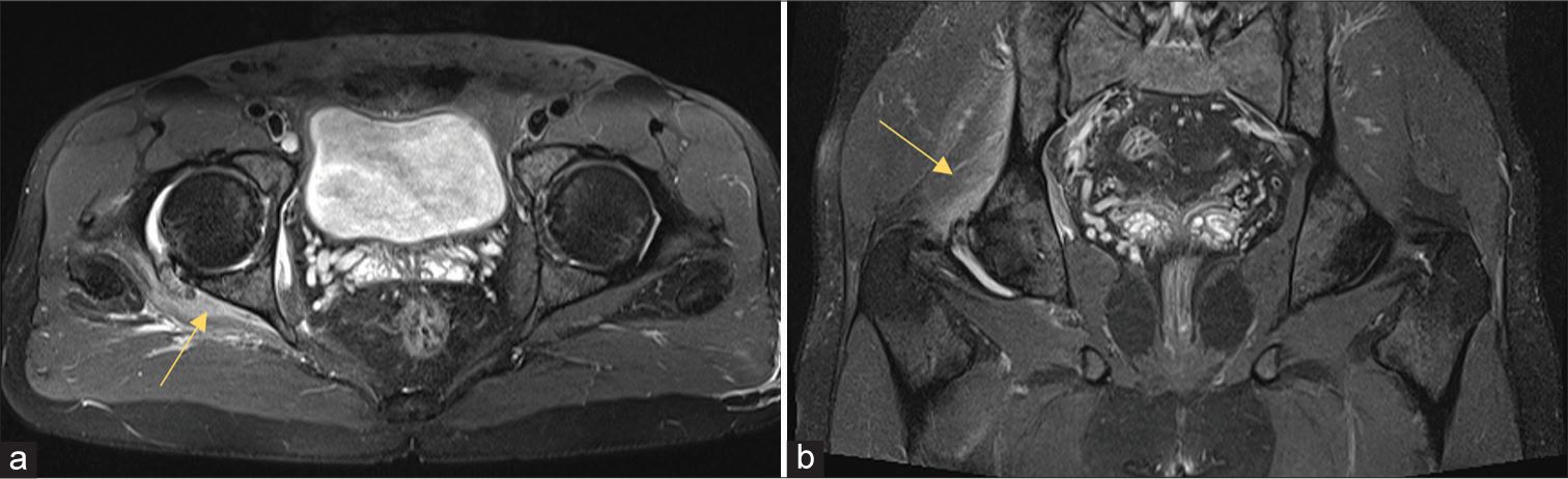
Diagnosis is confirmed through X-ray, ultrasound, or MRI showing the characteristic radio-opaque deposits. Calcium hydroxyapatite deposition appears as bright areas on plain film X-ray, distinguishing it from other soft tissue pathologies.
Erosion and Deposition: Geological Processes
Erosion and deposition are the two sides of the sediment transport cycle. Erosion removes rock and soil material from its original location through weathering, water flow, wind, glacier movement, or gravitational processes. Deposition occurs when the transporting agent loses enough energy to drop its sediment load — a river slows as it enters a lake, a glacier melts, wind velocity drops, and material settles.
Erosion and deposition work together to shape every landscape feature:
- River deltas form through deposition of eroded upstream sediment at the river’s mouth
- Beaches result from wave erosion of headlands and deposition of sand in lower-energy bays
- Glacial moraines are deposition ridges of material carried and dropped by glaciers
- Sand dunes form through wind erosion and deposition in arid and coastal environments
Calcium Hydroxyapatite Deposition: Treatment Options
Treatment for calcium hydroxyapatite deposition disease progresses from conservative to interventional depending on symptom severity:
- Conservative: NSAIDs for pain management, ice, rest from aggravating activities, and physical therapy after the acute phase to restore range of motion
- Corticosteroid injection: Ultrasound-guided injection into the bursa or peritendinous area reduces acute inflammation
- Needling (barbotage): Ultrasound-guided puncture and aspiration of the deposit, which disrupts the crystal mass and can accelerate resorption
- Extracorporeal shock wave therapy (ESWT): Non-invasive treatment that uses acoustic waves to break up deposits; evidence supports its effectiveness for chronic cases
- Surgical removal: Arthroscopic removal of deposits is reserved for cases that fail all conservative and minimally invasive treatments
Gadolinium Deposition Disease Symptoms
Gadolinium deposition disease symptoms represent an emerging area of medical concern following evidence that gadolinium-based contrast agents (GBCAs) used in MRI scanning can be retained in brain tissue, bone, and other organs — even in patients with normal kidney function. Traditional teaching held that GBCAs cleared completely from the body, but retention in brain nuclei (particularly the dentate nucleus and globus pallidus) has been documented through MRI signal changes and tissue analysis.
Reported gadolinium deposition disease symptoms include persistent headache, cognitive difficulties, bone and joint pain, vision changes, and skin sensations including burning, tingling, or tightening. The condition is distinguished from gadolinium toxicity (nephrogenic systemic fibrosis, a severe complication in patients with kidney failure) and from the documented signal changes on MRI, which are asymptomatic. Gadolinium deposition disease as a clinical entity is still debated in the medical community, with some specialists accepting it and others requiring more evidence before recognizing it as a defined diagnosis.